Sally K. Norton

Vitality Coach, Speaker & Health Consultant

  • Home
  • About
    • Interviews and Talks
  • Symptoms
  • Relief
    • Nourishment
    • Results
  • Science
    • Oxalate Basics
  • Books
    • Data Companion
  • Support
    • Upcoming Events
    • Support Groups
    • Shop / Downloads
    • Sign-up for News and Updates
    • Speaking and Presentations
  • Recipes
    • Fundamentals
    • Sauces and Condiments
    • Beverages
    • Finger Foods
    • Soups
    • Salads
    • Side Dishes
    • Meats and Seafood
    • Treats
  • Blog
    • Table of Contents
  • Contact
    • Share Your Success Story!
    • Coaching and Consulting
    • Speaking
    • Webmaster
    • Privacy Policy

April 9, 2017 by Sally K Norton

Sloppiness of Standard Practices in Science

Copyright: teamarbeit / 123RF Stock Photo

The public is unaware of many problems in how science is conducted and the ramifications for patient health.

For example, poor handling and significant time lapse before pathologists see the fragile and perishable tissues they examine can yield unreliable, meaningless data. Richard Harris discusses such problems in modern biomedical science in a new book, Rigor Mortis: How Sloppy Science Creates Worthless Cures, Crushes Hope, and Wastes Billions. The book is long overdue! It is certainly an important topic in need of some light, and I’m looking forward to reading it. Thanks to Richard Harris for tackling this massive problem!

In a book excerpt on the NPR site, Harris describes how researchers finally revealed one of the hidden problems in pathology.  While evaluating a breast cancer test, researchers in a lab at the University of Rochester let breast tissue biopsy samples sit out for an hour or two before testing them. They didn’t realize that at room temperature, the sample decays! Folks: dead tissues decay, and decay changes what a pathologist can detect.  Harris writes:Rigor Mortis Book Cover

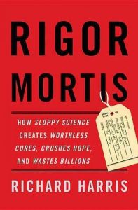
“And that was enough to degrade the sample and turn a positive result into a negative one. The molecule detected by the HER2 test breaks down at room temperature. “You can have the best test in the world and still get the wrong answer if you bugger up what you are testing,” Compton said.”

Oxalate Detection is a Problem for These Same Reasons Too

This problem of tissue degeneration is certainly true for oxalate crystals. These crystals are known to disappear from tissue samples within 2 hours of death, especially from the thyroid gland. Surgeons and their assistants lack training in the best methods for preserving biopsy samples and do not prioritize a speedy passage to the microscope. These processes are not priorities for biomedical institutions. Science and the public both suffer as a result.

I will write more soon about problems in tissue sampling and examination, focusing on overlooked oxalate deposits.  Unseen due to normal but faulty biomedical procedures, oxalate deposits in human tissues are more common than believed by researchers.

Link to NPR book excerpt:

http://www.npr.org/books/titles/522223284/rigor-mortis-how-sloppy-science-creates-worthless-cures-crushes-hope-and-wastes#excerpt

February 23, 2017 by Sally K Norton

Arterial Plaque Contains Oxalate

Did you know that oxalates can damage your heart and circulatory system, contributing to heart disease?

Our kidneys clean the blood. All of the oxalate that kidneys excrete every day gets there by traveling in our blood stream. The job of delivering toxic oxalate to the kidneys puts the arteries in harm’s way.

Researchers Don’t Expect Oxalate in Tissues

When oxalate deposits are found in tissues (other than the kidneys) doctors and researchers are surprised that the kidneys are working just fine. They believe that before oxalate collects in our bodies, the oxalate must first ruin the kidneys. This passage, below, from UCLA Pathologists Fishbein and Fishbein illustrates this conventional belief.

In a review of coronary arteries from 80 patients, we found 4 cases in which there were prominent oxalate deposits within the atherosclerotic plaques in coronary arteries. Oxalate deposits were also present in the media of arteries, the thyroid gland, and other organs, but not the kidneys, and the patients surprisingly did not have renal failure. (Fishbein and Fishbein 2009, p. 1315)

This expectation is based on the long-standing but unproven assertion that detectable (non-renal) oxalate deposits are a product of poor kidney function. In a previous report published by the journal Cardiovascular Pathology in 2008 they propose the new term “atherosclerotic oxalosis” to describe arterial plaque containing oxalate crystals.

This report was part of a study of coronary atherosclerosis in HIV infected patients, using atherosclerotic plaque samples from the National Neurologic AIDS Bank.

Whenever oxalate flows into the kidneys and other tissues in amounts beyond what they can handle (exceeding a capacity threshold), oxalate overload can occur. This can trigger accumulation of oxalate in the body. For some people this shows up as kidney deposits and kidney stones.  But oxalate can encounter, be stashed, and be left behind in any susceptible tissue in the body, not just the kidneys.

Other Forms of Oxalate are Usually Overlooked

Note the use of the term prominent in the comment quoted above. The researchers do not mention trace deposits, nano-crystals, ionic oxalate, and other chemical forms of oxalate in the examined tissues as a possibility. Unexplored, no doubt.

There is also no mention of the age of the specimens at the time of examination. We know that oxalate deposits rapidly fade in biopsied tissue and cadavers. (It would be interesting to see a study specific to arteries and arterial plaque to investigate the durability of less prominent oxalate deposits in plaque.) To reliably find oxalate, living tissues need to be examined within two hours of death or excision, but often are not. This may help to explain why oxalate in the arterial plaque is assumed to be rare, it can be hard to detect.

Calcium oxalate may be a factor in “hardening arteries”. And it may be more common than this research suggests, given the crude detection methods we use. Artery plaque oxalosis is most likely in people with metabolic stress such as HIV, metabolic syndrome, chronic inflammation, diabetes, insulin resistance, chemical injury, poly-pharmacy, auto-immune conditions, and so on. We don’t know how oxalate may harm “healthy” people with “perfect” arteries.

Over and over, science has demonstrated that oxalate can collect in any tissue in the body—and not necessarily due to kidney problems.

Reason enough to become oxalate aware!


Here is an update: an 2017 report of an Italian study looking at heart artery plaque in 229 people, obtained through a tissue bank. They did a special microanalysis in 41 plaque samples to differentiate between two types of calcifications: calcium oxalate vs hydroxapatite. 37% of the plaques were primarily calcium oxalate and 63% were hydroxyapatite. Oxalate calcifications were detected mainly in unstable plaques in 27% of cases, whereas HA tended to dominate unstable plaques 60% of the time.
Of course, unstable plaques are the ones that tend to break loose and block fine capillaries, restricting blood flow to brain or heart tissues thus causing non-hemorrhagic stroke or heart attacks . https://www.ncbi.nlm.nih.gov/pubmed/28739184

July 2, 2016 by Sally K Norton

Vitamin D Lamp: This little light of mine, finding time to let it shine.

Healthy Sun Solution with a Sun Lamp

Here in this video you can see my “electronic sun”. This easy-to-use vitamin D lamp has specialized UVB emitting bulbs intended to emulate natural sun. For most of us, brief sessions with such a lamp may improve our vitamin D status and general health. Making time in your busy life can be a challenge, however. Check out my own solution in the video to see how I fit this practice into my own schedule.

Read on to learn more about vitamin D and the advantages of the lamp.

Vitamin D

Deficiency of vitamin D is not only terrible for bone health, it increases your risk of almost any disease and lowers your life expectancy. Low D levels are epidemic these days. It is common in every age group, and especially common among dark-skinned people. This is because the natural way to “get” vitamin D is to make it with your own skin, when the sun shines.  The sun’s ultraviolet B (UVB) rays interact with cholesterol (7-DHC) in the skin to make precursors that your body can convert to vitamin D.  Skin pigment blocks UV penetration, protecting skin from UV damage, but limiting vitamin D production. Other factors that limit D production in skin include aging and sunscreen use.  The strength of the sun is also a factor, varying by time of day and season, and lower at higher latitudes.

Sunlight – We Need It.

Correcting vitamin D deficiency is an important aspect of health and healing. Vitamin D deficiency is associated with insulin disorders, autoimmune disorders, cancer, and even risk of infectious disease. Many experts recommend routine oral supplementation.  But there is some evidence of short-comings and side effects to this approach that we still do not understand well.

For those with inefficient vitamin D absorption, oral supplements do not correct the deficiency. For others who do absorb oral vitamin D, there are still drawbacks. Some suspect that daily consumption of D3 may encourage excessive absorption of calcium from the intestinal tract or movement of calcium out of the bones, which can create other problems.  To minimize these issues, it seems better to take D periodically in large doses, not in daily small doses.  But not everyone can tolerate pills and supplements at all:  such people especially need to generate vitamin D naturally from UVB rays (sunshine or sunlamp).

Practical Sun Exposure with a Lamp

Given the limitations of oral supplements, making the most of your skin’s ability to produce vitamin D is a good idea. But modern life makes it impractical to get regular sun exposure, even in the summer months. And UV radiation is insufficient in the northern U.S. during the winter months, November through February.  Many other variables influence how much UV light your body needs to make adequate vitamin D.

For pale skinned folks five minutes a few days a week might be enough. The darker your skin the longer the exposure needed. You can’t necessarily force your skin to make a lot of vitamin D from UVB, however. There are biological limits. So keep in mind that more time in the sun or under the lamp may not be better for you. Remember to have you doctor measure your vitamin D levels with blood testing.

Search on SallyKNorton.com

Shopping Cart

Number of items in cart: 0

  • Your cart is empty.
  • Total: $0.00
  • Checkout
Click to sign up for email list

Upcoming Events

  • MeatStock 2015: Oxalate Toxicity Talk

    May 18, 2025 @ 11:00 am - 11:40 am
    US Eastern Time
    See more details

  • Group Meeting and Presentation

    May 22, 2025 @ 2:00 pm - 4:00 pm
    US Eastern Time
    See more details

  • Group Meeting and Presentation

    June 5, 2025 @ 1:00 pm - 3:00 pm
    US Eastern Time
    See more details

Connect

  • Home
  • About
  • Table of Contents
  • Shop
  • Recipes
  • Support
  • Blog
  • Contact

Visit Sally’s Other Sites

  • YouTube
  • LinkedIn
  • Facebook
  • X
  • Instagram

Copyright © 2025 — Sally K. Norton • All rights reserved.


Loading Comments...